
TL;DR:
- Cardiac MRI segmentation is crucial for diagnosing cardiovascular diseases, but long-axis views are understudied.
- French research team introduces a robust augmentation strategy with ENet for automating segmentation.
- Hierarchical data augmentation improves segmentation accuracy, leveraging ENet’s efficiency.
- The methodology involves training on LAX 2-chamber and 4-chamber images with specific annotation rules.
- Significant enhancements were observed in segmentation quality and clinical metric estimation.
- ENet architecture shows promise for real-time whole-heart segmentation, despite minor precision losses.
Main AI News:
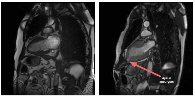
Addressing the imperative role of Cardiac Magnetic Resonance Imaging (CMRI) segmentation in diagnosing cardiovascular diseases, particularly ischemic heart conditions, is pivotal in the global battle against mortality. While CMRI stands as a beacon of precision in imaging anatomical regions with minimal risk, there exists a discernible gap in segmentation techniques, notably in long-axis (LAX) views, which are crucial for assessing atrial structures and diagnosing diseases affecting the heart’s apical region. To bridge this gap, state-of-the-art approaches have predominantly focused on short-axis (SAX) segmentation, leaving LAX views relatively understudied.
However, recent advancements such as the Ω-net method have heralded a shift in focus, acknowledging the significance of LAX views. By integrating statistical deformation models and data augmentation techniques like Generative Adversarial Networks (GANs), there lies a promising avenue for enhancing segmentation accuracy in CMRI. This not only aids in leveraging the unique advantages of LAX views for comprehensive cardiac imaging but also holds the potential for improving diagnosis and treatment strategies for cardiovascular ailments.
In line with this quest for precision, a recent paper by a French research team introduces a robust hierarchy-based augmentation strategy coupled with the Efficient-Net (ENet) architecture. This innovative approach aims to automate the segmentation of two-chamber and four-chamber Cine-MRI images, addressing the limitations of previous studies which primarily focused on SAX orientation. By leveraging the efficiency and effectiveness of ENet, the team endeavors to enhance segmentation accuracy in long-axis views, while delving into the impact of hierarchical data augmentation on segmentation quality.
The ENet architecture, renowned for its practicality and efficiency, emerges as a frontrunner in various medical imaging applications. In this study, researchers elaborate on its adaptation for cardiac Cine-MRI segmentation, with a specific focus on long-axis two- and four-chamber views. Unlike prior works concentrating solely on short-axis segmentation, this research delves into whole-heart segmentation in long-axis views, aiming to evaluate the efficacy of hierarchical data augmentation in enhancing segmentation accuracy.
The research unfolds with a meticulous approach toward producing anatomically accurate segmentation maps through a hierarchy-based augmentation strategy. Leveraging two datasets containing Cine-MRI LAX 2-chamber and 4-chamber images, the team trains the ENet architecture using the Adam optimizer on NVIDIA RTX 4500 GPU. Data augmentation is employed to refine segmentation accuracy through a combination loss function and a hierarchical procedure involving rotations, intensity alterations, and flipping. Evaluation metrics include the Dice coefficient, Hausdorff distance, and crucial clinical metrics like left ventricular volume and ejection fraction extrapolated from the segmentations.
The results showcase significant enhancements in segmentation quality, with notable improvements in Dice and Hausdorff distance. Additionally, clinical metric estimation, including Left Ventricular Ejection Fraction (LVEF), exhibits acceptable biases. This approach marks a significant stride forward in automated cardiac MRI segmentation, underlining the importance of considering long-axis representations for comprehensive cardiac evaluation.
Conclusion:
The research team presents an automated segmentation framework catering to the complexities of Cine-MRI LAX images, demonstrating robust results even in the face of anomalies and image degradation. The ENet architecture exhibits promise for whole-heart segmentation, offering compact sizes suitable for real-time applications. Despite minor precision losses near anatomical frontiers, the segmentation quality supports its clinical utility, paving the way for further optimization and advancements in cardiac MRI segmentation methodologies.