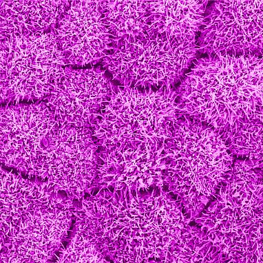
- Cutting-edge AI technology accelerates retinal imaging by 100 times and enhances image contrast by 3.5-fold.
- Dr. Johnny Tam’s team at the NIH pioneers the integration of adaptive optics (AO) with optical coherence tomography (OCT) for high-resolution retinal imaging.
- The development of the Parallel Discriminator Generative Adverbial Network (P-GAN) significantly reduces imaging acquisition and processing time while improving image quality.
- P-GAN outperforms traditional AI techniques in de-speckling retinal images, enabling precise visualization of cellular details.
- Dr. Tam envisions AI as an integral part of retinal imaging systems, revolutionizing diagnosis and treatment evaluation for retinal diseases.
Main AI News:
Cutting-edge artificial intelligence (AI) technology has transformed the landscape of retinal imaging, achieving remarkable milestones in speed and image quality enhancement. A recent study conducted by researchers at the National Institutes of Health has demonstrated that AI implementation in retinal imaging accelerates the process by a staggering 100 times, while simultaneously enhancing image contrast by 3.5-fold. These advancements hold significant promise in revolutionizing the assessment of age-related macular degeneration (AMD) and other retinal diseases, providing clinicians and researchers with an invaluable tool for diagnosis and treatment evaluation.
Dr. Johnny Tam, a leading figure in the Clinical and Translational Imaging Section at the NIH’s National Eye Institute, emphasized the critical role of AI in overcoming the time constraints associated with conventional retinal imaging techniques. His pioneering work focuses on integrating adaptive optics (AO) with optical coherence tomography (OCT), aiming to elevate the resolution and efficiency of imaging devices utilized in ophthalmic clinics worldwide. Tam’s analogy likens the transition from traditional OCT imaging to AO-enhanced OCT imaging as akin to upgrading from a distant balcony seat to a front-row perspective, enabling unparalleled insights into the intricate structures of the retina at a cellular level.
While the incorporation of AO into OCT undoubtedly enhances cellular visualization, it introduces challenges in image processing, particularly in addressing speckle interference. Speckle, akin to clouds obstructing aerial photography, poses a significant hurdle in obtaining clear and comprehensive images of retinal pigment epithelium (RPE) cells. Tam’s team tackled this obstacle head-on, devising a groundbreaking AI-based solution named the Parallel Discriminator Generative Adverbial Network (P-GAN). This deep learning algorithm, trained on a dataset comprising nearly 6,000 manually analyzed AO-OCT images of human RPE, excels in identifying and mitigating speckle-induced distortions, thereby facilitating the recovery of cellular details with unparalleled precision.
The efficacy of P-GAN was validated through rigorous testing, wherein it successfully de-speckled RPE images, surpassing the performance of conventional AI techniques. Dr. Vineeta Das, a postdoctoral fellow in the Clinical and Translational Imaging Section at NEI, underscored the transformative impact of P-GAN, estimating that it reduces imaging acquisition and processing time by a remarkable 100-fold while significantly enhancing image contrast. These findings herald a new era in retinal imaging, empowering clinicians with a potent tool for diagnosing and monitoring blinding retinal diseases with unprecedented efficiency and accuracy.
Dr. Tam envisions a future where AI seamlessly integrates into the fabric of retinal imaging systems, transcending its conventional role as a post-capture enhancement tool. By embracing AI as an integral component of the imaging process, rather than an afterthought, the field stands poised to unlock new frontiers in understanding the intricacies of retinal structure, function, and pathophysiology. The paradigm shift catalyzed by AI holds immense potential in democratizing access to advanced imaging technologies, heralding a future where precision medicine converges seamlessly with cutting-edge AI innovation.
Conclusion:
The integration of AI technology into retinal imaging represents a significant advancement in ophthalmology, offering unprecedented speed and accuracy in diagnosing and monitoring retinal diseases. This innovation holds immense potential for the market, driving the demand for AI-powered imaging solutions and paving the way for personalized and precise ophthalmic care.

