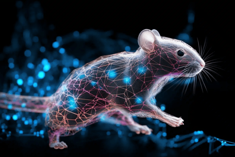
TL;DR:
- Recent technological advances enable the imaging of neurons in moving organisms.
- Computationally identifying and tracking neurons within dynamic, deforming brains has been a significant challenge.
- A collaborative effort between EPFL and Harvard introduces an AI-based method for neuron tracking.
- This method employs a convolutional neural network (CNN) to recognize and understand neuron patterns in images.
- Innovative ‘targeted augmentation’ reduces the need for extensive manual annotations, making the process more efficient.
- The method is versatile and capable of identifying neurons in various formats, including 3D volumes.
- Tested successfully on the roundworm Caenorhabditis elegans, revealing complex behaviors in interneurons.
- The team provides a user-friendly graphical interface for accessibility.
- This breakthrough significantly accelerates research in brain imaging and enhances our understanding of neural circuits and behaviors.
Main AI News:
In a groundbreaking development, recent strides in technology have enabled the imaging of neurons within organisms in motion. However, the critical challenge lies in computationally identifying and tracking these imaged neurons, especially when the brain undergoes movement and deformation within a flexible organism, such as a worm. Until now, the scientific community has grappled with the lack of suitable tools to tackle this intricate problem.
Enter a collaborative effort between scientists from EPFL and Harvard, who have unveiled a pioneering AI methodology for tracking neurons within dynamic and morphing organisms. This remarkable study, recently published in Nature Methods, was spearheaded by Sahand Jamal Rahi at EPFL’s School of Basic Sciences.
The core of this groundbreaking method revolves around a convolutional neural network (CNN), a potent form of AI meticulously trained to discern and decipher patterns within images. This process entails “convolution,” where the network scrutinizes small segments of the image, such as edges, colors, or shapes, and subsequently amalgamates this data to derive meaningful insights, thereby identifying objects or patterns.
However, a significant challenge arises when attempting to identify and track neurons amidst the constant fluctuations of an animal’s brain during a motion sequence. This is primarily due to the myriad body deformations the animal undergoes over time. The sheer diversity of the animal’s postures necessitates an extensive amount of manual annotations to train a CNN, a task that can be overwhelmingly daunting.
To address this monumental challenge, the researchers devised an enhanced CNN featuring a concept known as ‘targeted augmentation.’ This innovative technique autonomously generates reliable annotations for reference, utilizing a limited number of manual annotations. The result is that the CNN adeptly learns the intricate internal deformations of the brain, employing this knowledge to generate annotations for new postures. This groundbreaking approach significantly diminishes the necessity for manual annotation and cross-verification.
Moreover, this innovative methodology showcases remarkable versatility, capable of identifying neurons whether they appear in images as individual points or as intricate 3D volumes. The researchers put their creation to the test using the roundworm Caenorhabditis elegans, a favored model organism in the field of neuroscience, known for its 302 neurons.
Leveraging the enhanced CNN, scientists observed and measured the activities of certain interneurons within the worm, neurons responsible for bridging signals between others. Their findings were nothing short of extraordinary, revealing complex behaviors within these interneurons, including adaptive response patterns when subjected to various stimuli, such as periodic bursts of odors.
What’s more, the research team has generously made their CNN accessible to the scientific community, providing a user-friendly graphical interface that seamlessly incorporates targeted augmentation. This comprehensive pipeline streamlines the process, from initial manual annotation to the final proofreading stage.
Sahand Jamal Rahi, the driving force behind this transformative methodology, notes, “By significantly reducing the manual effort required for neuron segmentation and tracking, the new method increases analysis throughput three times compared to full manual annotation.”
This breakthrough holds the promise of expediting research in the realm of brain imaging, further enriching our understanding of neural circuits and behaviors. As the world of AI continues to revolutionize scientific exploration, this remarkable achievement paves the way for new horizons in neuroscience.
Conclusion:
The introduction of this groundbreaking AI method for tracking neurons within mobile organisms represents a significant leap forward in the field of neuroscience. By streamlining the process and reducing manual annotation efforts, it promises to accelerate research in brain imaging. This transformative advancement has the potential to deepen our understanding of neural circuits and behaviors, opening up new opportunities and applications in the market for neuroscience research tools and technologies.

