TL;DR:
- Osaka Metropolitan University researchers harness AI’s potential for heart health.
- AI application adeptly categorizes cardiac functions and detects valvular heart disease.
- Groundbreaking findings published in The Lancet Digital Health.
- Chest X-rays, a common diagnostic tool, reveal hidden cardiac insights.
- AI model trained on extensive dataset showcases impressive diagnostic accuracy.
- AI’s role is envisioned as enhancing diagnostics, bridging specialist gaps, aiding emergencies.
Main AI News:
In a realm often characterized by its cold, mechanical precision, Osaka Metropolitan University researchers have ignited a transformation that warms the heart—both literally and figuratively. Their pioneering utilization of artificial intelligence (AI) has birthed a new era of “heart-warning” support, as AI takes center stage in the realm of cardiac health.
Breaking down barriers at the crossroads of medical science and technology, the team’s groundbreaking AI application unveils a novel avenue for classifying cardiac functions and detecting valvular heart disease, marking a momentous leap towards enhanced patient outcomes. The prestigious pages of The Lancet Digital Health now bear witness to these epoch-making findings.
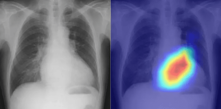
Valvular heart disease, a formidable foe of cardiovascular well-being, often eludes detection except through specialized echocardiography techniques. However, a shortage of adept technicians perpetuates diagnostic challenges. This is where the power of chest radiography—a common diagnostic tool mainly employed for pulmonary disorders—holds untapped potential. Surprisingly, these chest X-rays hold clues to cardiac function and maladies, a fact that remained hidden until now.
Within the corridors of countless medical institutions, chest X-rays are routine and swift, making them an easily accessible diagnostic tool. Dr. Daiju Ueda, the vanguard of this pioneering endeavor from the Department of Diagnostic and Interventional Radiology at Osaka Metropolitan University’s Graduate School of Medicine, envisioned these everyday scans as more than meets the eye. Could they serve as a reliable complement to echocardiography? Dr. Ueda’s team embarked on a journey to forge an AI model capable of skillfully classifying cardiac functions and valvular heart diseases, using chest radiographs as the enigmatic puzzle pieces.
Given the pitfalls of training AI on a solitary dataset, risking skewed outcomes, the team gathered an extensive 22,551 chest radiographs and corresponding echocardiograms from 16,946 patients across four facilities spanning the years 2013 to 2021. This rich tapestry of data served as the backdrop for the AI model’s training, linking the inputs of chest radiographs with the outputs of echocardiograms.
The fruits of this technological alchemy emerged as a precise classifier for six distinct valvular heart disease types. Evaluated by the esteemed Area Under the Curve (AUC) metric—a benchmark ranging from 0 to 1, where higher values denote superior performance—the AI model demonstrated AUC scores ranging from 0.83 to a remarkable 0.92. Notably, the AI’s prowess shone brightly in detecting left ventricular ejection fraction, a pivotal gauge of cardiac function, where it achieved an AUC of 0.92 at a 40% threshold.
Dr. Ueda expressed, “This journey has been one of endurance and significance. Our work stands to enhance diagnostic efficiency, especially in scenarios bereft of specialists, during nocturnal medical crises, and for patients averse to traditional echocardiography.”
Conclusion:
This remarkable stride in AI-driven cardiac assessment holds substantial promise for the medical technology market. The fusion of medical science and AI technology not only augments diagnostic accuracy but also addresses challenges posed by specialist shortages and time-sensitive scenarios. The successful application of AI in a critical domain like cardiac care underscores its potential to reshape the landscape of healthcare technology and services, creating opportunities for innovation and market growth.

