
TL;DR:
- Researchers in Australia have developed LoHiResGAN, an image-to-image translation model, to enhance low-field MRI scans.
- LoHiResGAN improves image quality while preserving diagnostic integrity, rivaling high-field MRI scans at 3 Tesla (3T).
- MRI’s non-invasive nature and radiation-free approach make it invaluable in medical science.
- Low-field MRI, specifically the 64 milliTesla (64mT) variant, offers cost-effective and portable solutions.
- Deep learning models like LoHiResGAN are used to translate low-field 64mT MRI scans into high-field 3T images.
- The study compared LoHiResGAN with other image-to-image translation models and found it excelled in image quality and brain morphometry measurements.
- GAN-based models, particularly LoHiResGAN, show promise in improving low-quality MRI scans.
- Challenges remain in accurately estimating brain measurements, highlighting areas for future improvement.
- LoHiResGAN’s success expands the scope of clinical diagnoses, particularly in regions without high-field MRI access.
Main AI News:
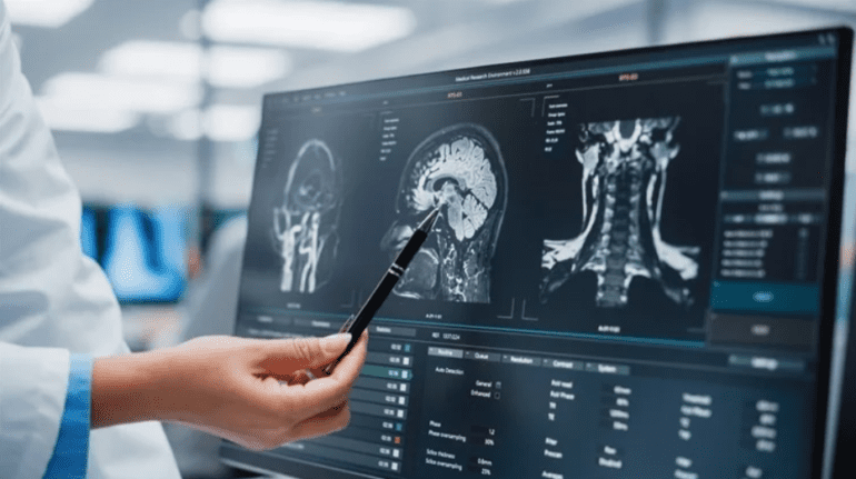
In a groundbreaking study recently featured in Scientific Reports, a team of Australian scientists has harnessed the power of deep learning to enhance low-field magnetic resonance imaging (MRI) scans. Their image-to-image translation model, LoHiResGAN, has not only preserved diagnostic integrity but also elevated image quality to rival that of high-field MRI scans operating at 3 Tesla (3T).
Magnetic resonance imaging has long been a cornerstone of medical science due to its non-invasive nature and its ability to provide high-contrast visuals of soft tissues and organs. Achieved through the amalgamation of radiofrequency pulses, strong magnetic fields, and cutting-edge computer algorithms, MRI scans can render images of various body regions, including the brain, joints, organs, and the vertebral column. Importantly, MRI’s radiation-free approach minimizes the risk of radiation-related complications.
Unlike their high-field counterparts, low-field MRI scanners, specifically the 64 milliTesla (64mT) variety, offer an economically efficient, compact, and portable solution. Despite a lower signal-to-noise ratio, low-field MRI has carved out a niche for itself, particularly in emergency settings and economically challenged or remote regions, making it an invaluable asset in the realm of medical imaging.
Recent research endeavors have zeroed in on the development of deep-learning-based models aimed at translating low-field 64mT MRI scans into high-field 3T images. In this study, a paired dataset comprising 64mT and 3T scans, with a focus on T1 weighted MRI to enhance fatty tissue signals and T2 weighted MRI to enhance water signals, served as the foundation for evaluating LoHiResGAN’s performance against other image-to-image translation models like CycleGAN, GANs, cGAN, and U-Net.
The study recruited 92 healthy participants who underwent scans using both 3T and 64mT MRI systems. Brain scans were meticulously analyzed, with morphometric measurements spanning 33 brain regions compared across images from 64mT, 3T, and synthetic 3T scans generated by the models.
Careful considerations were made when selecting the imaging sequences. The researchers opted for a two-dimensional T2-weighted turbo spin echo (TSE) sequence for the 3T MRI scans, ensuring efficient scanning while minimizing patient discomfort. Moreover, to ensure immediate clinical relevance, the researchers tailored their results to align with established clinical practices.
The preparation of the deep-learning model training data involved the use of a linear image resolution tool to co-register the 3T and 64mT scans. The dataset was subsequently divided randomly into three groups—training, validation, and testing—to ensure rigorous evaluation.
LoHiResGAN, the model proposed in this study, employs a Residual Neural Network (ResNet) component within a Generative Adversarial Networks (GAN) architecture. Its effectiveness was scrutinized by comparing its performance to models lacking ResNet components. Quantitative metrics, including the structural similarity index measure, normalized root-mean-squared error, perception-based image quality evaluator, and peak signal-to-noise ratio, were employed to assess the various image-to-image translation models’ performance.
The results unveiled a remarkable achievement by LoHiResGAN, as the synthetic 3T images it generated outperformed those produced by other models like CycleGAN, GANs, cGAN, and U-Net in terms of image quality. Additionally, LoHiResGAN yielded more consistent brain morphometry measurements across various brain regions when compared to the other models.
While all image-to-image translation models demonstrated enhanced signal-to-noise ratios and structural similarity index measures compared to low-field 64mT scans, GAN-based models, particularly LoHiResGAN, exhibited superior quantitative metrics over the U-Net model. These findings underscored the potential of GAN-based models in elevating the quality of low-quality MRI scans.
However, it is crucial to note that while low-field MRI scans offer logistical advantages, discrepancies may still arise, potentially affecting clinical diagnoses. Precise estimations of brain morphometric measurements, vital for diagnosing conditions like hydrocephalus, can be challenging. The study’s researchers also acknowledged certain limitations of deep-learning-based models, such as difficulties in accurately labeling white and grey matter in the brain, indicating areas for future improvement.
In summation, the findings of this study illuminate LoHiResGAN as a game-changer in the realm of low-field MRI imaging, significantly enhancing image quality while maintaining consistency in morphometric measurements across diverse brain regions. These breakthroughs open doors to expanding the scope of clinical diagnoses, especially in regions where high-field MRI scans may not be readily available. The future of medical imaging appears brighter than ever, thanks to the innovative fusion of deep learning and MRI technology.
Conclusion:
The development of LoHiResGAN represents a significant advancement in the field of medical imaging. Its ability to enhance low-field MRI scans while maintaining diagnostic accuracy has the potential to revolutionize clinical diagnoses, particularly in underserved regions with limited access to high-field MRI technology. This innovation opens new opportunities for the medical imaging market, with a growing demand for deep learning-based solutions to improve image quality and broaden the applications of MRI in healthcare.