
TL;DR:
- Traditional mitotic counting in breast cancer diagnosis is labor-intensive and prone to errors.
- Subjectivity in identifying hotspots and lack of standardization lead to discrepancies among pathologists.
- Digital pathology and AI integration offer a more accurate and efficient approach.
- AI algorithms standardize hotspot identification, improve consistency, and reduce pathologists’ workload.
- High-quality, diverse training data and ongoing validation are essential for AI tools’ effectiveness.
- The challenge of non-standardized data collection hampers AI adoption in pathology.
- AI integration in breast cancer diagnosis enhances precision, efficiency, and speed.
- The combination of AI and pathologists’ expertise creates a more responsive and resilient healthcare system.
- AI has the potential to revolutionize not only breast cancer diagnosis but also medical diagnostics as a whole.
Main AI News:
In the realm of breast cancer pathology, mitotic counting serves as a cornerstone in disease staging and diagnosis. Seasoned pathologists understand the critical role that precision plays in this step, but they are also acutely aware of the labor-intensive and error-prone nature of traditional methods. As pathology labs grapple with an increasing caseload, the demand for a more accurate and efficient approach has never been more pressing. Enter artificial intelligence (AI), a crucial ally that is significantly elevating the capabilities of pathologists in the realm of breast cancer diagnosis.
Traditional Mitotic Counting and Its Challenges
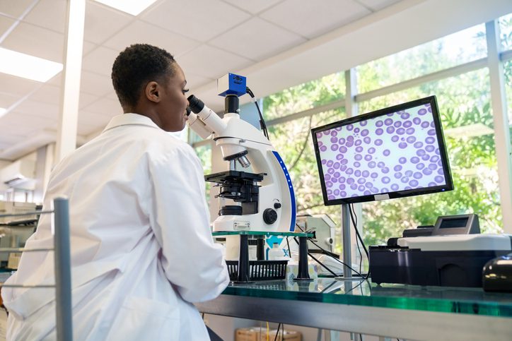
At the heart of breast cancer diagnostics lies mitotic counting, where pathologists meticulously inspect glass slides under the microscope. The task entails identifying a hotspot – an area rich in mitotic events – and manually tallying each occurrence. However, traditional mitotic counting faces a multitude of challenges that can jeopardize its reliability. Subjectivity in pinpointing the precise hotspot often leads to discrepancies among pathologists. In fact, recent research published in the Journal of Clinical Pathology reveals that pathologists frequently disagree on their observations, potentially leading to errors in cancer severity determination and treatment decisions. This is exacerbated by the tedious nature of counting mitoses, coupled with the mounting workload pressures.
Another significant drawback of conventional techniques lies in their lack of standardization. Microscopes vary in magnification and field areas, introducing further inconsistencies in the counting process. These disparities can have profound implications on patient diagnoses and prognoses, given that the mitotic index is a pivotal parameter in breast cancer grading.
The Emergence of Digital Pathology and AI Integration
The transition to digital pathology has marked a pivotal advancement in breast cancer diagnosis. High-resolution digital imaging of tissue slides provides pathologists with an unprecedentedly clear and comprehensive view of patient samples. The integration of digital tools, including automated measurements, area grids, and advanced annotation capabilities, enhances the accuracy and efficiency of the diagnostic process. However, it is the synergy between AI and these digital tools that heralds the most transformative shift.
AI algorithms, when overlaid onto digital pathology, introduce a new level of precision and efficiency. These sophisticated applications have been meticulously designed to surmount the challenges traditionally faced by pathologists. With AI, the subjective process of hotspot identification through human eyes and microscopes can be standardized, mitigating variability and ensuring consistency across diagnoses. AI systems systematically annotate each mitotic figure within these hotspots, leaving no crucial detail unexamined. Moreover, these tools can automatically compute mitotic counts across entire slides and within specific hotspots, substantially alleviating the workload of pathologists and expediting the diagnostic process.
Developing Effective AI Tools: Crucial Considerations
To integrate such AI into clinical practice, a foundation of high-quality, diverse training data is imperative. This guarantees that AI algorithms can adeptly recognize and analyze the spectrum of histological features encountered in various patient samples. Ongoing testing and validation by practicing pathologists are vital to maintaining the accuracy and clinical relevance of these AI systems. Furthermore, soliciting direct feedback from pathologists in the design and refinement of AI tools ensures that these systems are attuned to the real-world intricacies of the diagnostic process.
Scientific validity hinges not only on dataset size but also on statistical significance and demographic representation mirroring the broader population. The medical community has long grappled with non-standardized data collection, especially concerning racial and ethnic disparities, which often go unreported due to varying levels of documentation across health systems, insurance providers, and public health records. This challenge remains a significant hurdle for most datasets undergoing FDA review, explaining why, out of over 500 AI algorithms approved by the FDA, only one is sanctioned for clinical use in pathology.
Embracing a New Era in Breast Cancer Diagnostics
As breast cancer diagnostics evolve, the integration of AI opens up a realm of possibilities. Pathologists armed with AI tools are already delivering more precise, efficient, and expeditious diagnoses. This marks a significant stride in a field where the speed and precision of AI complement the nuanced expertise of experienced pathologists, creating a healthcare landscape that is not only more responsive but also more resilient. As AI continues to mature and integrate seamlessly into the clinical workflow, its potential to revolutionize not only breast cancer diagnosis but also the broader spectrum of medical diagnostics promises to usher in an era of better outcomes for every patient.
Conclusion:
The integration of AI into breast cancer diagnosis represents a significant advancement, addressing the limitations of traditional methods. This innovation not only improves accuracy and efficiency but also holds promise for transforming the broader landscape of medical diagnostics. As AI continues to evolve, it will play a pivotal role in ensuring better outcomes for patients and reshaping the healthcare market by enhancing diagnostic capabilities.