
TL;DR:
- Scientists at Johns Hopkins University have harnessed AI to visualize and monitor changes in the strength of synapses in living organisms.
- The technique offers insights into how synapses evolve with learning, age, trauma, and disease in human brains.
- By leveraging machine learning, researchers can enhance the quality of images and track individual synapses in multiday experiments.
- The study provides a novel approach to analyzing synapse-level changes, paving the way for a better understanding of brain function.
- Collaboration between neuroscience and biomedical engineering experts led to this breakthrough, showcasing the power of interdisciplinary research.
Main AI News:
Cutting-edge artificial intelligence (AI) has revolutionized the field of neuroscience by enabling researchers to witness and monitor the dynamic transformations occurring within neural synapses. In a groundbreaking development, scientists from Johns Hopkins University have harnessed the potential of AI to create a pioneering technique for visualizing and tracking alterations in the strength of synapses, the crucial junctions that facilitate communication between nerve cells in the brain. Detailed in a recent publication in Nature Methods, this technique has the potential to unlock a deeper understanding of how these connections evolve in the human brain, influenced by factors such as learning, aging, trauma, and disease.
Dr. Dwight Bergles, the esteemed Diana Sylvestre and Charles Homcy Professor in the Solomon H. Snyder Department of Neuroscience at the Johns Hopkins University School of Medicine, elucidates the significance of this breakthrough, stating, “If you want to learn more about how an orchestra plays, you have to watch individual players over time, and this new method does that for synapses in the brains of living animals.” By capturing the intricate dance of synapses within living organisms, scientists can now embark on a transformative journey to unravel the mysteries of the brain.
The study, co-authored by Dr. Bergles and his esteemed colleagues Dr. Adam Charles, Dr. Jeremias Sulam, and Dr. Richard Huganir, represents a remarkable collaboration between the fields of neuroscience and biomedical engineering. All four researchers are esteemed members of the renowned Kavli Neuroscience Discovery Institute at Johns Hopkins University. At the core of this research lies the understanding that nerve cells transmit information by exchanging chemical messages at synapses, and various life experiences, such as exposure to new environments and the acquisition of skills, are believed to induce changes in these synapses, ultimately facilitating learning and memory formation. Yet, comprehending these minute changes across the trillions of synapses within the human brain has posed an immense challenge—one that this novel technique is poised to conquer.
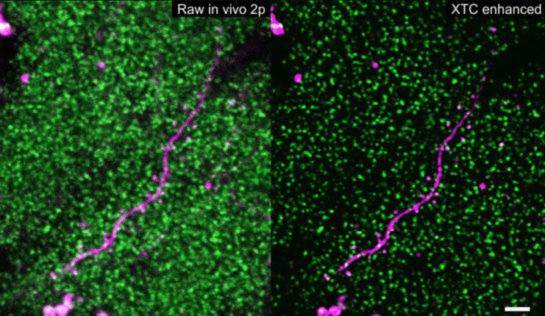
Traditionally, scientists have grappled with the daunting task of visualizing the dynamic chemistry of synaptic messaging. The high density and diminutive size of synapses render them incredibly challenging to observe even with state-of-the-art microscopes, necessitating a breakthrough solution. Dr. Charles explains the predicament, stating, “We needed to go from challenging, blurry, noisy imaging data to extract the signal portions we need to see.” Enter machine learning—the computational framework that has revolutionized numerous biomedical imaging domains. Leveraging the power of machine learning, the team of researchers devised an approach to enhance the quality of images encompassing thousands of synapses. This remarkable tool surpasses human speed, automating the detection process, but first, it must be “trained” to recognize what high-quality images of synapses should resemble.
In their groundbreaking experiments, the researchers worked with genetically modified mice in which the glutamate receptors—critical chemical sensors at synapses—glowed with a fluorescent green hue when exposed to light. The amount of fluorescence emitted by each receptor serves as an indicator of the number and strength of synapses. However, capturing clear images of individual clusters of these receptors within the intact brain proved to be a formidable challenge. To surmount this obstacle, the team employed a two-pronged approach. They trained a machine learning algorithm using high-quality images derived from brain slices, allowing them to produce enhanced images with significantly improved resolution. Furthermore, they captured low-quality images that closely resembled those obtained from live animals, thereby ensuring a more accurate representation of synapses in their natural environment.
This ingenious cross-modality data collection framework paved the way for the development of an enhancement algorithm capable of transforming low-quality images into high-resolution representations. By employing this technique, researchers can now discern and track individual synapses, numbering in the thousands, throughout multi-day experiments conducted in the intact brains of living mice. The ability to explore the dynamic changes occurring within synapses over time is invaluable in unraveling the intricacies of neural networks.
To trace the temporal changes in receptors within living mice, the team employed microscopy to capture repeated images of the same synapses over several weeks. After establishing baseline images, the animals were placed in an environment enriched with novel stimuli, including new sights, smells, and tactile sensations, for a brief five-minute period. Subsequently, the researchers imaged the same region of the brain every other day to observe how these new stimuli influenced the number of glutamate receptors at synapses. The findings were remarkable: even this simple change in environment elicited a spectrum of alterations in fluorescence across synapses in the cerebral cortex. These variations indicated connections where the strength increased, as well as connections where it diminished, with a notable bias toward strengthening in animals exposed to the novel environment. The implications of these discoveries extend far beyond this specific experiment, offering a glimpse into the vast potential for analyzing synapse-level changes in diverse contexts.
The success of this groundbreaking research lies in the close collaboration between scientists with diverse areas of expertise, spanning molecular biology to artificial intelligence. The interdisciplinary environment fostered at the Kavli Neuroscience Discovery Institute has been instrumental in nurturing such collaborative efforts. Dr. Bergles emphasizes the significance of these partnerships, stating, “We are really excited to see how and where the rest of the scientific community will take this.” Indeed, the possibilities are boundless. As this machine learning approach continues to unveil the secrets hidden within synaptic connections, it holds immense promise for illuminating the synaptic changes that occur in various diseases and injury contexts, including prominent conditions such as Alzheimer’s disease.
Dr. Sulam concludes with an air of anticipation, “We are really excited to see how and where the rest of the scientific community will take this.” The potential for future advancements in neuroscience appears limitless as researchers embark on a transformative journey, driven by the marriage of artificial intelligence and cutting-edge experimental techniques.
Conclusion:
The groundbreaking use of AI to uncover and monitor synaptic changes in the brain signifies a significant leap forward in neuroscience. This technique not only provides valuable insights into the intricate workings of the brain but also opens up new avenues for research in various domains, including learning, aging, trauma, and disease. The successful collaboration between experts from different fields highlights the importance of interdisciplinary approaches in driving scientific progress. From a market perspective, this breakthrough has the potential to spur advancements in neurology, pharmaceuticals, and brain-related therapies, with implications for areas such as Alzheimer’s disease research and the development of targeted treatments. The integration of AI and neuroscience holds immense promise, and its impact on the market is expected to be transformative in the coming years.