
- Harvard researchers and Google collaborators publish a detailed 3D map of a small segment of the human brain in Science.
- Imaging of a cubic millimeter of tissue generated 1.4 petabytes of data.
- Artificial intelligence utilized to stitch microscope images together, reconstructing the sample in 3D.
- Project spans a decade, involving meticulous reconstruction of neural networks from excised brain tissue.
- Insights reveal glia outnumber neurons 2:1, oligodendrocytes dominate, and rare potent axonal inputs exist.
- Full paper accessible in Science, with dataset and viewer provided for public exploration.
Main AI News:
In the realm of scientific advancement, while Large Language Models (LLMs) have been taking the spotlight, various AI techniques are quietly permeating numerous fields. Take, for instance, a recent publication in Science by Harvard researchers, alongside collaborators from Google, unveiling a meticulous 3D map of a minute section of the human brain, rendering astonishing detail. This feat of imaging, capturing approximately a cubic millimeter of tissue, amassed a staggering 1.4 petabytes of data.
Nature magazine encapsulates this groundbreaking endeavor, quoting Viren Jain, a neuroscientist at Google, who remarks, “It’s a little bit humbling. How are we ever going to really come to terms with all this complexity?” Jain and his team leveraged artificial intelligence to piece together microscope images, reconstructing the entire sample in 3D. Reflecting on the experience, Jain reminisces, “I remember this moment, going into the map and looking at one individual synapse from this woman’s brain, and then zooming out into these other millions of pixels. It felt sort of spiritual.”
This monumental undertaking spans a decade of relentless effort. Initiated approximately ten years ago, a minute portion of human brain tissue found its way to Dr. Jeffrey Lichtman’s laboratory at Harvard. Extracted during a procedure to alleviate seizures in an epilepsy patient, the tissue underwent meticulous reconstruction by Lichtman’s team. Employing a $6 million device, the sample was sliced into paper-thin sections, and through electron microscopy, intricate neural networks were painstakingly recreated.
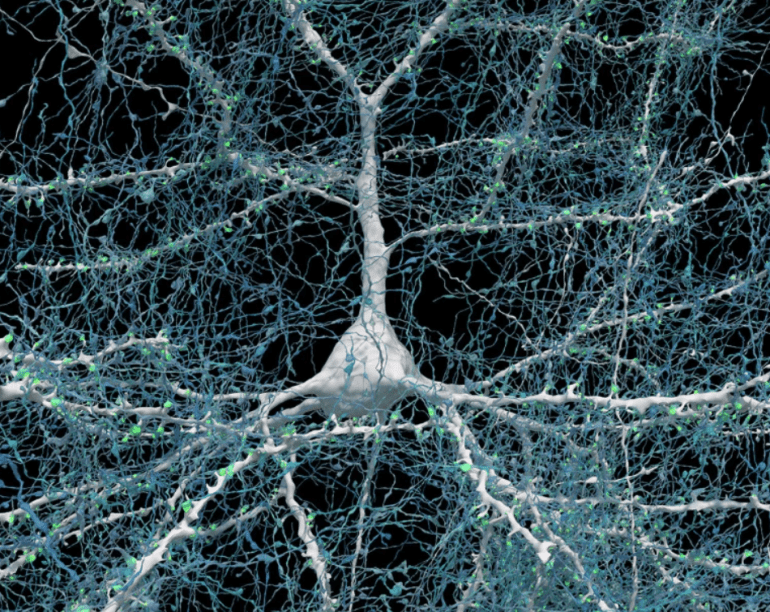
The images captured are nothing short of remarkable. Providing an abstract, the magnitude of the project becomes apparent: “Presented here is a computationally intensive reconstruction of the ultrastructure of a cubic millimeter of human temporal cortex… It contains about 57,000 cells, about 230 millimeters of blood vessels, and about 150 million synapses and comprises 1.4 petabytes.” This analysis uncovers intriguing insights, revealing, among other findings, the prevalence of glia over neurons, the dominance of oligodendrocytes, and the classification of deep layer excitatory neurons based on dendritic orientation. Moreover, it sheds light on the existence of rare potent axonal inputs.
While the complete paper resides within the annals of Science, Google and the research team extend public access to the dataset and Neurglancer viewer, inviting individuals to explore this invaluable resource.
Conclusion:
The groundbreaking 3D mapping of a segment of the human brain not only underscores the advancements in imaging technology and artificial intelligence but also opens avenues for further research into the complexities of the human brain. This breakthrough signifies a significant leap in understanding neural networks and holds promise for advancements in neuroscience and medical diagnostics. Companies in the medical imaging and AI sectors should take note of the potential applications and opportunities that arise from such advancements in brain mapping technology.